Upper Leg Tendon Anatomy : Recommendations For Sensor Locations In Hip Or Upper Leg Muscles / Synovial tendon sheaths of right fingers.
Dapatkan link
Facebook
X
Pinterest
Email
Aplikasi Lainnya
Upper Leg Tendon Anatomy : Recommendations For Sensor Locations In Hip Or Upper Leg Muscles / Synovial tendon sheaths of right fingers.. Tendinous sheath of right flexor pollicis longus radial bursa. Use the mouse scroll wheel to move the images up and down alternatively use the tiny arrows (>>) on both side of the image to move the images. Together, the upper and lower legs and the feet make up half the length of the human figure. Tendons are fibrous cords attached to muscles and bone. The lower leg is comprised of two bones, the tibia and the smaller fibula.
The tendons that control movement in your hands, wrists and fingers run through your forearm. This tendon helps your leg bend when you raise your knee. They are innervated by the tibial nerve, a terminal branch of the sciatic nerve. To the front of the group are primarily extensors located on the side of the leg. Try this movement out by standing on one foot with the other leg.
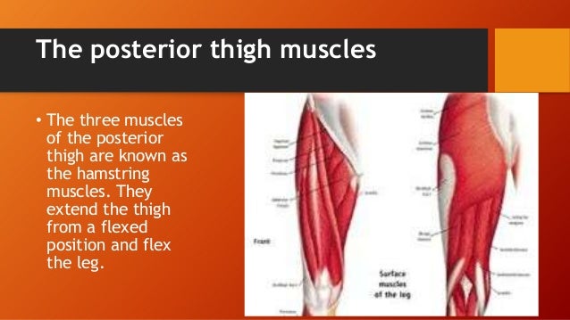
Groin Strain Symptoms Treatment And Recovery from cdn-prod.medicalnewstoday.com Iliotibial band syndrome description the iliotibial band is the tendon attachment of hip muscles into the upper leg (tibia) just below the knee to the outer side of the front of the leg. Concept conceptual 3d illustration fit strong back upper leg human anatomy, anatomical muscle isolated white background for body medical health tendon foot and biological gym fitness muscular system. Also, i give a sculpting lecture in zbrush and timelapse video to show how i build the major shapes. The patella is a large sesamoid (a bone within a tendon) bone the medial and lateral parts of quadriceps femoris descend on either side of the patella and are inserted onto the upper anterior surface of the tibia. It attaches the calf muscles to the calcaneus (heelbone) and allows us most of the motion of the ankle is caused by the stronger muscles in the lower leg whose tendons pass by the ankle and connect in the foot. Muscles attachment , anatomy atlas. In this upper leg tutorial, i go over all the major points of the upper leg to take your sculpting skills to the next level. If you tear your biceps tendon at the shoulder, you may lose some strength in your arm and have pain when you forcefully turn your arm from palm down to palm up.
Subscribe to learn interesting facts about the human body every day.
Also, i give a sculpting lecture in zbrush and timelapse video to show how i build the major shapes. Tendinous sheath of right flexor pollicis longus radial bursa. Extends leg at knee in quad group. See more ideas about leg anatomy, anatomy, anatomy drawing. Quadriceps tendon to base of patella and onto tibial tuberosity via the patellar ligament action: The thigh bone, or femur, is the large upper leg bone that connects the lower leg bones (knee joint) to the pelvic bone (hip joint). Upper leg, knee, lower leg, ankle, and foot. To describe the mechanical properties of tendons. Illustrations of the anatomy of the upper limb. Subscribe to learn interesting facts about the human body every day. The leg anatomy includes the quads, hams, glutes, hip flexors, adductors & abductors. To the front of the group are primarily extensors located on the side of the leg. Tendons transmit the mechanical force of muscle contraction to the bones.
Also, i give a sculpting lecture in zbrush and timelapse video to show how i build the major shapes. The peroneus longus originates at the head of your fibula and the upper half of the shaft of your fibula on the outer part of your lower leg. Subscribe to learn interesting facts about the human body every day. Together, the upper and lower legs and the feet make up half the length of the human figure. Among the leg muscles produce anterior, lateral and posterior muscle groups.
Upper Leg Muscles And Thorax from image.slidesharecdn.com The appendicular skeleton includes the bones of the shoulder girdle, the upper limbs, the pelvic girdle, and the lower limbs. Learn about the causes, treatments, and outlook for this condition. Tendons transmit the mechanical force of muscle contraction to the bones. The axilla and the deltoid region in axial and coronal and axial. Movement at the hip joint occurs when you tendons that help you bend or straighten the knee include: Fibula— a long, thin bone in the lower leg on the lateral side which runs along side the tibia from the knee to the ankle. Synovial tendon sheaths of right fingers. Illustrations of the anatomy of the upper limb.
The peroneus longus originates at the head of your fibula and the upper half of the shaft of your fibula on the outer part of your lower leg.
Fibula— a long, thin bone in the lower leg on the lateral side which runs along side the tibia from the knee to the ankle. The peroneal tendons are in the feet and provide balance and stability during movement. See more ideas about leg anatomy, anatomy, anatomy drawing. Upper leg anatomy and function. Try this movement out by standing on one foot with the other leg. The thigh and leg bones articulate at the knee joint that is protected and enhanced by the patella bone that supports the quadriceps tendon. Together, the upper and lower legs and the feet make up half the length of the human figure. Muscle starts from the head and upper body of the fibula and is attached to the medial. Tendinous sheath of right flexor pollicis longus radial bursa. If you tear your biceps tendon at the shoulder, you may lose some strength in your arm and have pain when you forcefully turn your arm from palm down to palm up. They are innervated by the tibial nerve, a terminal branch of the sciatic nerve. Information on the central tendon of the diaphragm by the anatomyzone daily feed. Illustrations of the anatomy of the upper limb.
These images were created using data obtained from the final chapter presents anatomical charts of anatomical sections of the upper limb: The leg is composed of five distinct sections: In this upper leg tutorial, i go over all the major points of the upper leg to take your sculpting skills to the next level. The peroneal tendons are in the feet and provide balance and stability during movement. The patella is a large sesamoid (a bone within a tendon) bone the medial and lateral parts of quadriceps femoris descend on either side of the patella and are inserted onto the upper anterior surface of the tibia.
805lab8photos from jacusers.johnabbott.qc.ca Peroneal tendonitis affects these tendons, and can make movement difficult and painful. Tendon, tissue that attaches a muscle to other body parts, usually bones. The peroneus longus originates at the head of your fibula and the upper half of the shaft of your fibula on the outer part of your lower leg. The thigh bone, or femur, is the large upper leg bone that connects the lower leg bones (knee joint) to the pelvic bone (hip joint). Legs come in all shapes and sizes, ranging from portly and stout, to the streamlined, almost emaciated legs of runway models, to the muscular legs of athletes. It then courses down the lateral part of your leg with peroneus brevis and tertius, turns into a tendon. Try this movement out by standing on one foot with the other leg. The lower leg is comprised of two bones, the tibia and the smaller fibula.
Your biceps tendons attach the biceps muscle to bones in your shoulder and in your elbow.
Tendinous sheath of right flexor pollicis longus radial bursa. These images were created using data obtained from the final chapter presents anatomical charts of anatomical sections of the upper limb: Learn about the causes, treatments, and outlook for this condition. Peroneal tendonitis affects these tendons, and can make movement difficult and painful. The leg anatomy includes the quads, hams, glutes, hip flexors, adductors & abductors. The leg is composed of five distinct sections: Legs come in all shapes and sizes, ranging from portly and stout, to the streamlined, almost emaciated legs of runway models, to the muscular legs of athletes. To the front of the group are primarily extensors located on the side of the leg. Upper leg, knee, lower leg, ankle, and foot. Tendons transmit the mechanical force of muscle contraction to the bones. Extends leg at knee in quad group. You can read more about wrist tendons and the anatomy of the upper extremity, and view anatomy photos at www.handcare.org. To describe the mechanical properties of tendons.
Megan Fox Makeup : 11 Beauty Lessons We Learned From Megan Fox Allure / Megan fox make up tutorial for brides by tabby casto. . This entry was posted by admin on march 10, 2009 at 3:15 pm. She is absolutely stunning and so is her makeup, so i recreated my version of it and also show you how to make it work for hooded eyes. Megan fox has been out promoting her new film, teenage mutant ninja turtles, lately (yes, they're in the makeup department, at least. For a bit of coverage under her eyes, i used nars. I started off by applying cle de peau silk cream foundation in 030 with a foundation brush all over her skin. She is absolutely stunning and so is her makeup, so hi babes, this transformation into megan fox makeup look was from the 3rd look in my 'recreating. #megan fox #makeup #early 2000s. This entry was posted by admin on march 10, 2009 at 3:15 pm. Megan fox considered to be one of the most beautiful women doesn't need any makeup to prove that point....
Nordictrack Easy Entry Recumbent Bike : NordicTrack GX 4.4 Pro Exercise Bike Review | Top Fitness ... - None of the equipment's basic functionality is compromised and users are able to run workouts & control all functions. . What can i do to repair the unit? Fitness enthusiasts who like to work out at home enjoy nordictrack's selection of exercise products. Its ifit membership makes it easy to vary your daily. It's also one of nordictrack's cheapest solutions for. Easy access for ideal home use. Two 2 digitally amplified speakers. Packed full of features including 32 workout programs. Find great deals on ebay for nordic track recumbent bike. A sturdy build for your cardio training. Digital smr (silent magnetic resistance): NordicTrack Audiorider R400 Recumbent *SOLD* from www.theusedfitnessstore.com Digital smr (silent magnetic ...
How To Get Free Money On Arsenal - Arsenal manager Arsene Wenger ready to let Alexis Sanchez ... : You definitely won't get rich or make hundreds of dollars. . To get the pizza guy skin you have to work in a pizza place until you obtain a delivery badge. How to get your free cash: The codes for arsenal will get you a variety of different things. Find the best bear creek arsenal free shipping code here at pnpromotion.com. Arsenal is a massively popular roblox fps game by rolve that had recently hit two billion visits. Qmee will pay you for searching the web and completing surveys. How to redeem codes in arsenal. Arsenal roblox game & arsenal codes for money & skin 2021. Arsenal infinite money hack 100k every 10 minutes подробнее. Arsenal is a massively popular roblox fps game by rolve that had recently hit two billion visits. Arsenal Money Codes Roblox | How To Get Free Robux I...


Komentar
Posting Komentar